IMACEL Case Studies
Support Case#03
Pathology specimen analysis ~Abnormality screening and virtual staining~
Challenge
Pathological examinations in drug safety assessments are traditionally performed by direct microscopic observation. However, this process is time and labour intensive because toxicologists must thoroughly examine the entire surface of every slice generated.
In addition, as the diversification of drug discovery modalities increases, so does the likelihood of encountering new types of lesions that have not been previously observed. This raises concerns that AI models trained solely on the appearance of known lesions will be limited in practical implementation.
Furthermore, the effects of artifacts generated during the specimen preparation process and inter-facility variations must also be taken into consideration.
LPIXEL’s Solution – AI Technology and Impact
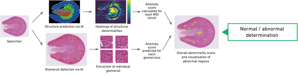
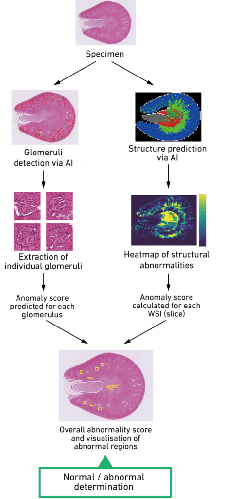
AI-based pathological specimen screening
Two specialized AI models are trained on a wide range of normal kidney tissue images, enabling them to detect deviations from normal (abnormalities) in new specimens:
- Detection of tubular abnormalities visible at lower magnification (capturing disruption of the overall tissue structure)
- Detection of glomerular abnormalities (Identifying minute changes)
By combining the outputs of these two dedicated models, the AI system determines whether a specimen is overall normal or abnormal.
As this approach evaluates abnormality based on the degree of deviation from normal tissue, rather than relying solely on recognizing specific, predefined lesions, this method is expected to be effective even for previously unseen, novel toxicities.
We have successfully achieved detection of both tubular and glomerular lesions, along with visualizations that illustrate the extent of deviation from normal tissue structure.
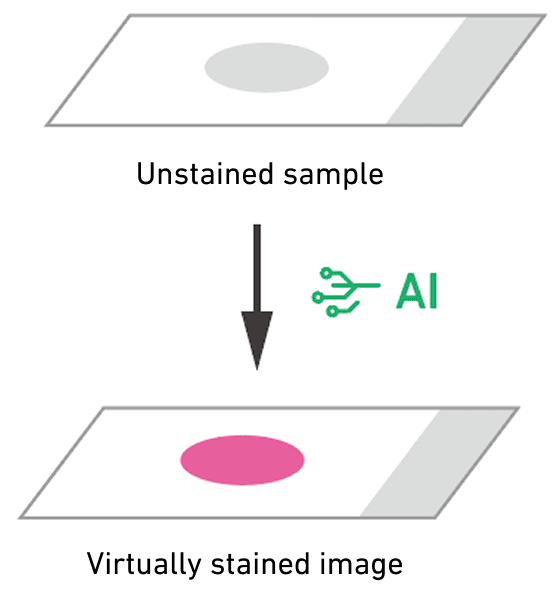
Virtual Staining
By generating virtually stained images from unstained samples using AI, inter-facility variability in staining can be eliminated to achieve consistent, uniform image analysis. This approach also allows (virtual) staining to be applied to tissue specimens reserved for genetic analysis without degrading the original material.
As another example, by generating immunostained images from existing HE slides, researchers can investigate specific biological mechanisms by leveraging the large archive of HE images already available. This also allows researchers to pinpoint regions of interest before performing the actual immunostaining.

AI-Based Histopathological Lesion Assessment for Safety Evaluation in Rat Kidney and Testis
— Joint presentation with Daiichi Sankyo Co., Ltd.
(The 41st Annual Meeting of the Japanese Society of Toxicology, 2025)
第41回日本毒性病理学会学術集会「Artificial intelligenceを用いたラット腎臓における糸球体および尿細管の病理組織学的病変の安全性評価」(第一三共株式会社との共同発表・2025年)
Support Case#01
Phenotypic Screening
~Identifying candidate compounds via cell morphology analysis~
Support Case#02
Automated fluorescent multiplex immunohistochemistry image analysis
~Applied to anticancer drug research and development (R&D) and spatial analysis of the tumor microenvironment.~
Support Case#03
Pathology specimen analysis
~Abnormality screening and virtual staining~
Support Case#04
Automated Light-Sheet Microscopy Image Analysis
~Automatic Brain Region Segmentation and Improved Efficiency in Perivascular Environment Analysis~
Support Case#05
Novel approaches to animal behaviour analysis using AI
~Advanced behavioural insights and precise quantification~
Support Case#06
Automated Micronucleus Test Evaluation
~Provided as an automatic analysis service~
Support Case#07
Automation of Chromosomal Abnormality Detection
~Applications in dose assessment and genotoxicity testing following radiation overexposure~
Support Case#08
Ice Crystal Analysis in the Freeze-Drying Process
~Optimizing Manufacturing Processes with AI~
Support Case#09
Lab Automation Support
~Integrating image-analysis AI into automated medium-scale synthesis systems~