IMACEL Case Studies
Support Case#02
Automated fluorescent multiplex immunohistochemistry image analysis ~Applied to anticancer drug research and development (R&D) and spatial analysis of the tumor microenvironment.~
Challenge
Fluorescent multiplex Immunohistochemistry (IHC, a technique that uses fluorescent dye-labeled antibodies to simultaneously detect multiple molecules expressed in cancer cells, immune cells, and other tissues on a pathological specimen) enables the measurement of expression levels of multiple molecules at the single-cell level, allowing for detailed cell classification.
Daiichi Sankyo uses LPIXEL’s AI technology to automate the analysis of fluorescent immunostained tissue images focusing on Antibody-Drug Conjugate (ADC) target molecules and immune cells.
Traditionally, skilled researchers relied on conventional (non-AI-based) analysis software that required manually setting and verifying analysis parameters through visual inspection. This presented significant challenges, including lengthy analysis times and results that were dependent on the analyst’s skill and experience.
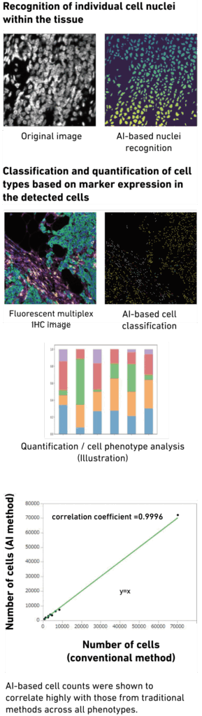
LPIXEL’s Solution – AI Technology and Impact
The automation of fluorescent multiplex IHC image analysis was achieved through the following two-step process:
Recognition of individual cell nuclei within the tissue
Classification and quantification of cell types based on marker expression in the detected cells
Analyses were conducted across multiple cancer types, including breast, colorectal, and pancreatic cancers.
Results demonstrated that the AI achieved cell nucleus recognition accuracy and quantitative cell classification performance comparable to conventional methods.
Going forward, LPIXEL will expand the range of cancer types supported and integrate analyses with other experimental data, such as spatial omics, to realize quantitative spatial analysis of the tumor microenvironment.

https://www.daiichisankyo.co.jp/files/news/pressrelease/pdf/202207/20220720_J2.pdf
https://www.daiichisankyo.co.jp/files/investors/library/materials/2024/ST_Day_2024_final_J.pdf
https://www.daiichisankyo.co.jp/files/news/pressrelease/pdf/202207/20220720_J2.pdf
https://www.daiichisankyo.co.jp/files/investors/library/materials/2024/ST_Day_2024_final_J.pdf
Support Case#01
Phenotypic Screening
~Identifying candidate compounds via cell morphology analysis~
Support Case#02
Automated fluorescent multiplex immunohistochemistry image analysis
~Applied to anticancer drug research and development (R&D) and spatial analysis of the tumor microenvironment.~
Support Case#03
Pathology specimen analysis
~Abnormality screening and virtual staining~
Support Case#04
Automated Light-Sheet Microscopy Image Analysis
~Automatic Brain Region Segmentation and Improved Efficiency in Perivascular Environment Analysis~
Support Case#05
Novel approaches to animal behaviour analysis using AI
~Advanced behavioural insights and precise quantification~
Support Case#06
Automated Micronucleus Test Evaluation
~Provided as an automatic analysis service~
Support Case#07
Automation of Chromosomal Abnormality Detection
~Applications in dose assessment and genotoxicity testing following radiation overexposure~
Support Case#08
Ice Crystal Analysis in the Freeze-Drying Process
~Optimizing Manufacturing Processes with AI~
Support Case#09
Lab Automation Support
~Integrating image-analysis AI into automated medium-scale synthesis systems~